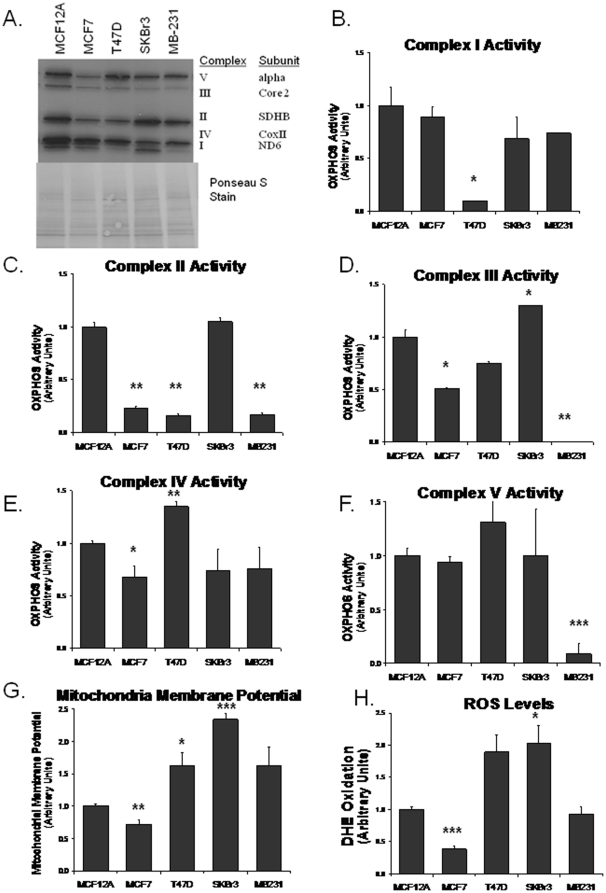
Figure 1. Changes in OXPHOS subunit gene expression and activity in breast cancer cells.
A. Western blot analysis of a representative subunit from each complex. Ponceau S staining serves as a loading control. Oxidative phosphorylation enzyme activities were measured on isolated mitochondrial protein. B. Complex I activity was measured as the rotenone inhabitable rate of NADH oxidation. C. Complex II activity was measured by the succinate induced rate of reduction of DCIP. D. Complex III activity was measured as the rate reduction of cytochrome c (III) when stimulated with CoQ2H2. E. Complex IV activity was measured as the rate of cytochrome c (II) oxidation. F. Complex V activity was measured by the oxidation of NADH in the presence of pyruvate kinase/lactic dehydrogenase and PEP. G. Mitochondrial membrane potential was measured by TMRE fluorescence. H. Intracellular ROS was measured by DHE oxidation. Data are expressed as the mean ratio to MCF12A + 1 SEM, * p<0.05, ** p<0.005, *** p<0.0005.

