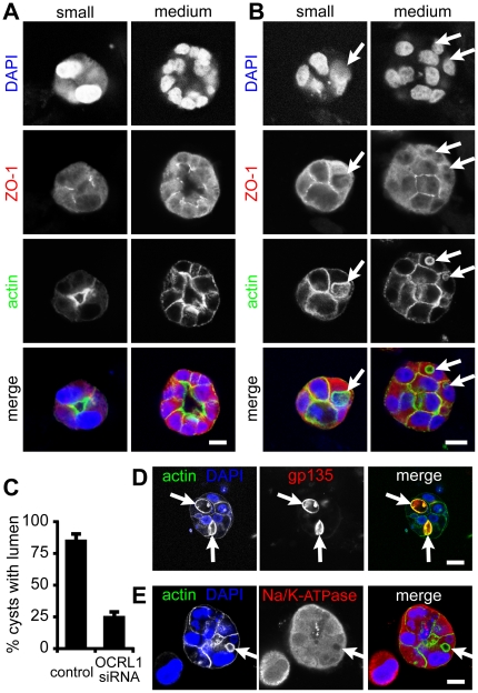
Figure 7. OCRL1 depletion interferes with lumen formation in MDCK cyst morphogenesis.
(A) MDCK cells were treated twice with control, non-targeting siRNA for 72 hours prior to being seeded into collagen/matrigel and allowed to proliferate for 4 days. Cells were fixed with 3% paraformaldehyde, permeabilized, blocked and processed for staining with DAPI, anti-ZO-1 and phalloidin (blue, red and green in merge respectively). Images (single confocal sections) are from either small clumps (2 to 4 nuclei in widest cross-section), or medium-sized clumps (approx. 10–15 nuclei in widest cross-section). Size bars = 10 µm. (B) As in A, but with four duplexes targeting OCRL1. Arrows indicate actin-rich spherical, intracellular vacuoles and the positions these occupy in alternately stained images. (C) Phalloidin images from two independent experiments were analysed for lumen formation. A total of 84 cysts were counted. 85% of control siRNA treated cell clumps were hollow cysts with lumens. In comparison, lumen formation was only seen in 25% of clumps of cell with silenced OCRL1. Bars show range from 2 experiments; t-test of these data showed the difference was significant (p<0.01). (D and E) MDCK cells were treated as in B. and stained with DAPI (blue) and phalloidin (left-hand panels, green in the merge), and antibodies either to gp135 (podocalyxin) or to sodium/potassium ATPase (right-hand panels, red in the merge). Scale bars are 10 µm.

