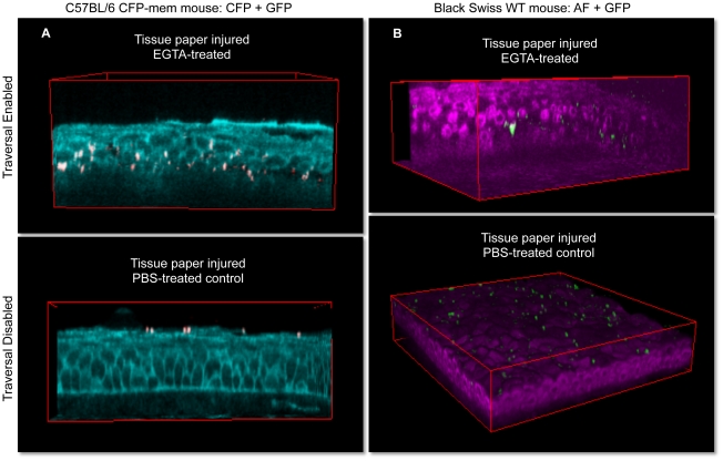
Figure 2. 3D imaging of bacterially challenged eyeballs.
All eyeballs were tissue paper blotted to enable bacterial adherence to the corneal epithelium. Susceptibility to traversal by adherent bacteria was then toggled on/off by use/omission of EGTA treatment following the blotting procedure. Upper images (EGTA used to toggle on traversal) show deep bacterial traversal 6 h after bacterial challenge in EGTA-treated corneas. Lower images were PBS-treated controls (EGTA step omitted) and showed bacterial adherence to the surface without subsequent bacterial traversal. Panel (a) shows confocal technique using mice with CFP-tagged cell membranes (cyan), and panel (b) shows two-photon images of cellular NAD(P)H autofluorescence (magenta) in transgenic (C57BL/6 background) or wild-type (Black Swiss background) mouse corneas respectively. Field size: 189 µm × 189 µm.

