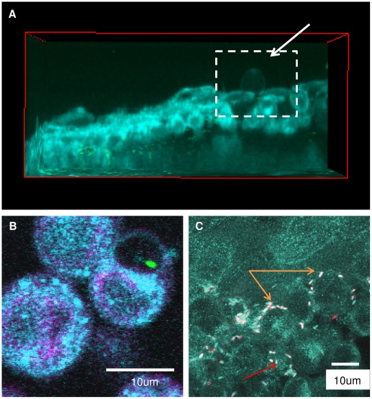
Figure 3. Temporal and spatial tracking of EGTA-enabled bacterial traversal.
Traversal was enabled in the corneal epithelium of transgenic mice expressing CFP-tagged cell membranes (cyan). Panels (a, b) show bacterial-induced membrane bleb formation ex vivo visualized as spherical membrane projections (arrow) extending away from the epithelial cells. A representative view in the xz plane is shown in (a), and a higher magnification imaging revealing a bleb-confined bacterium is shown in (b). In (c), bacteria can be seen located between cells (orange arrows) where some were motile (fast-moving GFP-bacteria, red; slower-moving GFP-bacteria, white = captured twice in both CFP and GFP channels). Other bacteria in this image appeared to be in the cytoplasm (red arrow).

