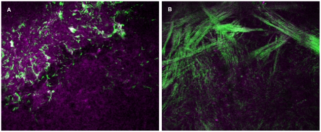
Figure 4. Imaging of bacteria and cells within opaque infected mouse corneas.
Panel (a) shows NAD(P)H autofluorescence (magenta) of corneal epithelial cells and bacteria (green) detected from optically opaque (infected) mouse eyes 24 h post-scratch and inoculation. In panel (b), bacteria (green) within the stroma of the cornea were found to orient themselves along the pattern of the collagen fibrils. Field size: 189 µm × 189 µm.

