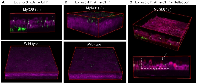
Figure 6. Impact of MyD88 deficiency on defense against epithelial traversal by bacteria after blotting.
Panel (a) shows two-photon images of cellular NAD(P)H autofluorescence (magenta) of MyD88 (−/−) (top) and wild-type (bottom) mouse corneas. Deep bacterial traversal (GFP, green) of corneal epithelium was detected in MyD88 (−/−) but not wild-type mice 8 h after bacterial challenge ex vivo. Only adherent bacteria without epithelial traversal were found in tissue paper-blotted wild-type corneas. Panel (b) shows an earlier (4 h) time-point at which a relatively larger number of bacteria were found adhering to the MyD88 (−/−) corneal epithelium as compared to the wild-type. In panel (c), merged image (fuchsia) of reflection (red) and autofluorescence (magenta) is shown. Reflection confocal methodology (red) can be used to capture cells not visualized by autofluorescence (i.e. dying or dead cells) to monitor cell viability during bacterial traversal. This method confirmed that penetrated bacteria at the later (8 h) time point were overlaid with epithelial cells.

