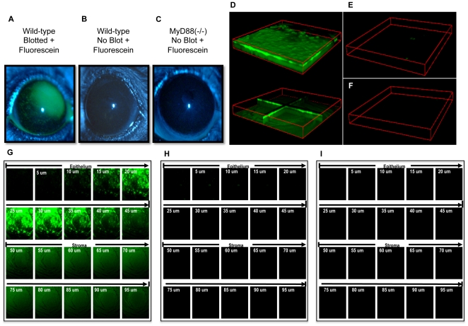
Figure 8. MyD88 corneas do not label with fluorescein suggesting tight junctions are intact ex vivo and in vivo.
Fluorescein was added to tissue paper blotted or normal healthy (non-blotted) mouse corneas to access cell-cell junction integrity. Extent of corneal staining was examined using a slit lamp (a-c) and confocal microscopy (d–i). Panels (a, d, g) show extensive fluorescein staining (green) in blotted wild-type (C57BL/6) corneas, but not non-blotted wild-type (b, e, h) or MyD88 (−/−) (c, f, i) mouse corneas. Z-stack confocal images (1272 µm × 1272 µm × 100 µm) are presented as 3D block view (d, top), 3D orthoslice view (d, bottom), and 2D x-y view (g) for blotted and fluorescein stained wild-type mouse cornea. In parallel, 3D block views and 2D x-y views of confocal images are shown for normal wild-type (e, h) and MyD88 (−/−) (f, i) mouse corneas. These data show that epithelial tight junctions are intact in MyD88 knockout corneas prior to bacterial exposure.

