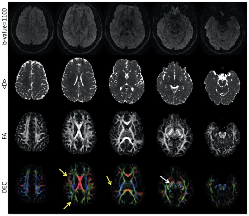
FIG. 5.

Five slices from a whole brain SSEPI DTI acquisition at 1.7mm isotropic voxel size. Diffusion-weighted images (top row), calculated <D> maps (second row), calculated FA (third row), and DEC maps (bottom row) are displayed. The yellow arrows point to sub-cortical white matter, while the white arrow points to the optic tract on the left side.
