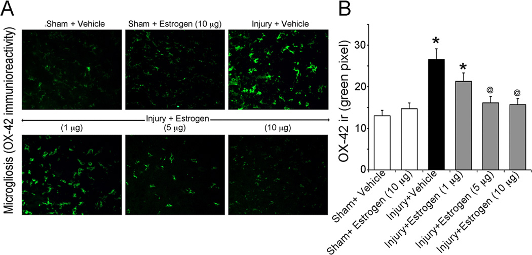
Figure 1.
Assessment of microgliosis in acute SCI animals. Spinal cord injured animals were treated with low doses of estrogen (1, 5, 10 µg/kg/day via osmotic pump) or vehicle and compared to sham surgery alone. Microgliosis was measured by OX-42 immunoreactivty in thin (5 µm) frozen sections. (A) Representative images (200X) are shown from the ventral horn of caudal penumbra spinal cord tissue. (B) Bar diagram quantifying OX-42 immunoreactivity. Fluorescence intensity was measured in a per field manner with 3 fields per animal. *P < 0.05 compared to sham + vehicle; @P < 0.05 compared to injury + vehicle (n ≥ 3 animals).

