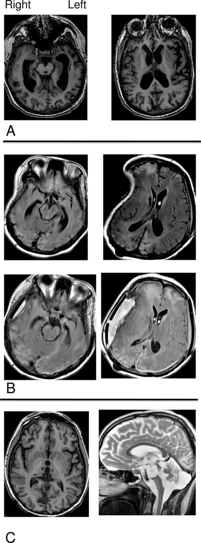
Figure 1. Brain MRIs showing major features of structural damage in the three patient subjects (PSs).
A. PS 1: T1-weighted MRI shows diffuse atrophy. B. PS 2: T2 FLAIR MRI shows focal injuries to frontal and occipital lobes and distortion from craniectomy on right (visit 1, top), and right occipital and bifrontal injuries, and fluid collection under cranioplasty site on right (visit 2, bottom). C. PS 3: T1-weighted axial image shows bithalamic and right medial temporo-occipital lobe strokes with minimal cerebral atrophy; T2-weighted sagittal image shows loss of majority of midline pons and midbrain.

