
Figure 2.
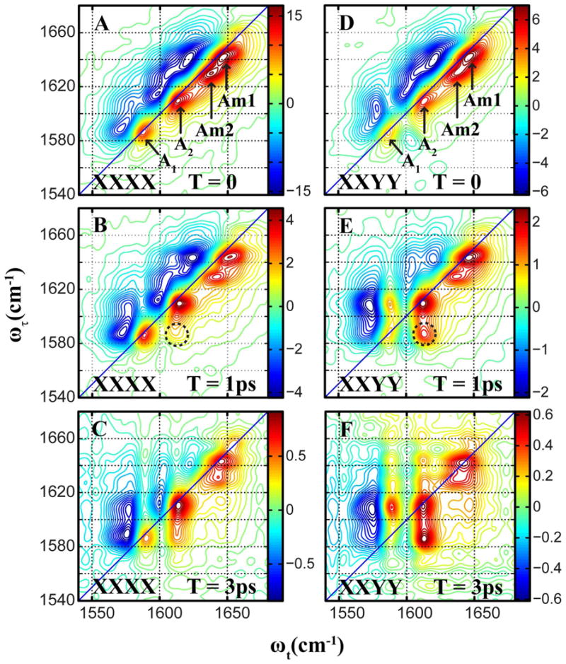
Absorptive 2D-IR spectra of arginine dipeptide in D2O for waiting times 0, 1ps and 3ps. The spectra for the XXXX polarization scheme are plotted on the left column, and those for the XXYY polarization scheme are plotted on the right column. The two Amide-I modes are labeled Am1 and Am2. The cross peak region is shown in dotted circles.