Figure 2.
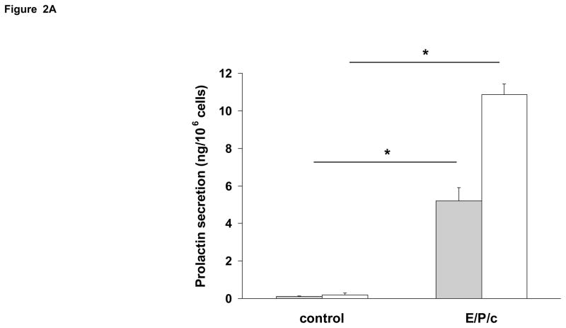
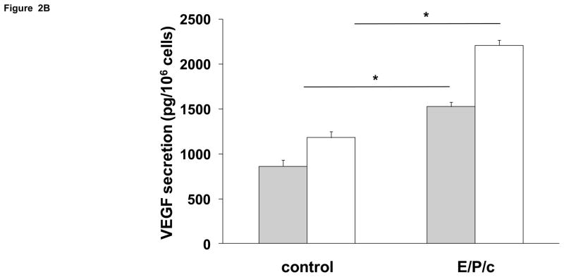
Time course of biochemical differentiation of ESC exposed to E/P/c. (A) Prolactin and (B) VEGF secretion both increased following hormone stimulation in vitro. Grey histograms indicate protein secretion after 4 d incubation without (control) and with hormones (E/P/c) and white histograms show the protein concentrations after 8 d incubation (*P<0.05, t-test).


