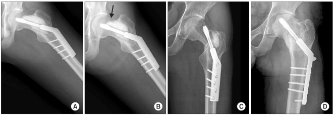
Fig. 3.
The anteroposterior radiographs of a 21-year-old man with fibrous dysplasia of his left femur neck show (A) the three months postoperative view after the curettage and injectable calcium sulfate grafting with prophylactic fixation. (B) Six months postoperatively, he complained of continuous pain on his left hip, and the radiograph showed the cortical breakage on the superior aspect of the femur neck (arrow). (C) Ten months postoperatively, complete fracture occurred in spite of activity restriction. Note that the calcium sulfate is not completely resorbed. (D) The final radiograph after curettage of the remnant calcium sulfate and internal fixation with autogenous bone grafting.

