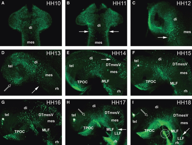
Fig. 1.

Time series of axon tract development in the chick embryonic brain. (A, B) Dorsal views of the brain; (C–I) lateral views of the brain. (A) HH10: no staining of neurones in the rostral brain. (B) HH11: neurones are located rostral to the DMB, within the diencephalon (arrows). (C) HH12: the number of MLF neurones has increased (arrow). (D) HH13: the first TPOC neurones are labelled in the rostral hypothalamus (open arrow). In the pretectum, MLF neurones are projecting axons caudally (filled arrow). (E) HH14: the MLF axon tract is projecting along the basal mesencephalon, and the TPOC neurones have started to project their axons. DTmesV neurones have started differentiating in the dorsal mesencephalon (arrow). (F) HH15: TPOC axons have reached the pretectum, forming a joint VLT with the MLF. Olfactory placode neurones are labelled in the telencephalic area (asterisk). (G) HH16: the number of MLF, TPOC and DTmesV neurones has increased, and their axons have projected further. (H) HH17: there are neurones located in the alar diencephalon projecting axons ventrally towards the TPOC (open arrow). These neurones are projecting dorsally towards the VLT. In the mesencephalon DTmesV axons are pioneering the LLF (filled arrow). (I) HH18: more alar neurones are present in the diencephalon (open arrow). The LLF is pioneered from the DTmesV (arrow). Circle shows the trigeminal nerve. (F–I) Asterisks mark the olfactory placode. di, diencephalon; DTmesV, descending tract of the mesencephalic nucleus of the trigeminal nerve; LLF, lateral longitudinal fascicle; mes, mesencephalon; MLF, medial longitudinal fascicle; rh; rhombencephalon; tel, telencephalon; TPOC, tract of the postoptic commissure.
