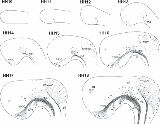
Fig. 2.

Development of axon tracts in the rostral chick brain. Sketches of the developing neurones and tracts in the rostral brain of chick embryos during the first 72 h of development. Note that the drawings are not representative of the absolute number of neurones present. HH10: no differentiated neurones detectable. HH11: the first MLF neurones differentiate. HH12: increased number of MLF neurones, but no neurones elsewhere yet. HH13: MLF neurones project axons caudally. TPOC neurones are visible in the rostral hypothalamus, and the first DTmesV neurones start appearing at the dorsal midline of the mesencephalon. HH14: MLF neurones are located in three subnuclei. Axons of the TPOC project towards the DMB. HH15: TPOC and MLF form a continuous, VLT. Axons of the DTmesV reach towards the alar-basal boundary in the mesencephalon. The first neurones in the olfactory placode region are detectable. HH16: MTT neurones are detected in the caudal hypothalamus, and their axons join the VLT system. The DTmesV axons turn caudally towards the MHB. HH17: all major tracts increase in complexity. Additional neurones are found scattered in the rostral diencephalon, and neurones also differentiate in the mesencephalic alar plate. HH18: the TPC becomes apparent in the caudal pretectum. VLT and DTmesV dominate the organisation of the rostral brain, but the rising number of additional neurones and axons progressively mask the early axon scaffold. The oculomotor nerve and its neurones are also prominent features of the early basal mesencephalon, but as peripheral nerve are not considered further in this study. III, oculomotor nerve; DTmesV, descending tract of the mesencephalic nucleus of the trigeminal nerve; LLF, lateral longitudinal fascicle; MLF, medial longitudinal fascicle; MTT mamillo-tegmental tract; TB, tecto-bulbar axons; TN, terminal nerve; TPC, tract of the posterior commissure; TPOC, tract of the postoptic commissure.
