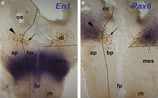
Fig. 3.

Patterning and MLF neurone location. In situ hybridisation (blue) for patterning genes En1 (A) and Pax6 (B) combined with immunohistochemistry (brown) for neurones detected with the Tuj1 antibody in HH12 flat-mounted chick brains. Black lines indicate the DMB as defined by the caudal limit of the Pax6 expression domain, and the alar plate-basal plate boundary as defined by the ventral limit of the Pax6 expression domain. (A) En1 is expressed in the mesencephalon. All of the neurones are located rostral to the En1 expression domain in alar (arrowhead) and basal plate (arrow). (B) Pax6 is expressed in the alar plate of the diencephalon. Neurones are located both in the Pax6-positive alar plate (filled arrowhead) and in the corresponding basal plate (arrow). Some neurones are just caudal to the Pax6 expression domain (open arrowheads). ap; alar plate; bp, basal plate; di, diencephalon; fp, floor plate; mes, mesencephalon; os, optic stalk; rh, rhombencephalon.
