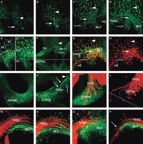
Fig. 4.

Development of the VLT. (A–D) Formation of the MLF axon tract. (A) HH11: first neurones are detectable in the alar plate (arrowhead) and basal plate (arrow) of the caudal diencephalon. (B) HH13: the number of neurones in the diencephalon has increased and some basal neurones have begun projecting axons caudally (arrow). (C) HH14: neurones in alar plate (arrowhead) and basal plate (arrows) of the diencephalon project axons caudally into the MLF, which runs along the floor of the mesencephalon. Some scattered neurones in the basal mesencephalon also project into the VLT (open arrowhead). (D) HH15: the MLF neurones are organised into three distinct populations in the alar plate (dorsal population, arrowhead), in line with the MLF (central, filled arrow) and ventral to the MLF (rostroventral, open arrow). (E) Detail of the three MLF neurone populations in a HH15 chick embryo. The alar population is spread across p1 and p2. In the basal plate, the central population is mainly located in p1, while the rostroventral population is in p2. E1, E2 and E3 are higher magnifications of each neurone population. (E1) The dorsal neurones have large cell bodies and are dispersed across the alar plate. They initially project their axons ventrally before turning caudally. (E2) The central population of neurones is very dense and intermingled with the MLF axons. (E3) Neurones in the rostroventral population have smaller cell bodies. They project axons first at an angle dorsal-caudally before joining the MLF. (F) Overview of the longitudinal tract in a HH15 chick embryo. TPOC (open arrowhead) and MLF form a seemingly continuous tract system running along the basal plate of the prosomeres and the mesencephalon. The three populations of MLF neurones are indicated (filled arrowhead: dorsal population; filled arrow: central population; open arrow: rostroventral population). Line indicates ventral midline. (G,H) The developing MTT in HH16 chick embryo. Double-labelling of the early axon scaffold with Tuj1 (green) and HuC/D (red). MTT neurones (open arrowhead) are located rostral to the MLF neurones project their axons caudally with the TPOC axon tract. The three populations of MLF neurones are indicated (filled arrowhead: dorsal population; filled arrow: central population; open arrow: rostroventral population). (H) Only the red HuC/D staining labelling only the neuronal cell bodies. A neurone-free gap separates the MTT neurones (open arrowhead) from the most rostral MLF neurones in p2 (open arrowhead). (I–P) Formation of the TPOC axon tract. (I) HH13: the first TPOC neurones differentiate in the basal plate of the rostral secondary prosencephalon, ventral to the optic stalk. (J) HH15: the TPOC neurones have started to project caudally towards the DMB (arrows). The TPOC neurones are clearly separated from the MTT neurones in the caudal secondary prosencephalon (open arrow). There are also neurones located rostral to the nMLF, which are projecting axons back towards the nTPOC (arrowhead). (K) HH17: retrograde labelling of the TPOC (DiI, red) combined with Tuj1 staining (green). The nTPOC is now a very dense population of neurones. Retrograde labelled neurones are scattered across the whole population of neurones. (L) HH18: retrograde labelling of TPOC axons from the mesencephalon. Most of the labelled neurones are densely packed in the ipsilateral nTPOC, but some contralateral neurones are also labelled. Line indicates anterior midline. (M–P) Relative location of MLF and TPOC. (M) HH17; (N) HH18. Retrograde labelling of the MLF (DiO, green) and anterograde labelling of the TPOC (DiI, red). The two axon tracts are running close together, but the TPOC axons are projecting just dorsal to the MLF. (O) HH17: DiI labelling of the TPOC (DiI, red) combined with Tuj1 (green) staining. The TPOC axons project well dorsal to the rostroventral population of MLF neurones and also dorsal to most of the central population. (P) HH19: retrograde labelling of TPOC (DiI, red) and MLF (DiO, green) from the rostral rhombencephalon. Both dyes were injected into the basal plate, but DiO was injected slightly more ventrally. The DiI-labelled axons can be traced back to the rostral hypothalamus (arrow), while DiO labelling depicts the three populations of MLF neurones, indicating that TPOC (dorsal) and MLF (ventral) axons remain mostly separated also in their caudal projection. MLF, medial longitudinal fascicle; nTPOC, nucleus of the TPOC; p1, prosomere 1; p2, prosomere 2; TPOC, tract of the postoptic commissure.
