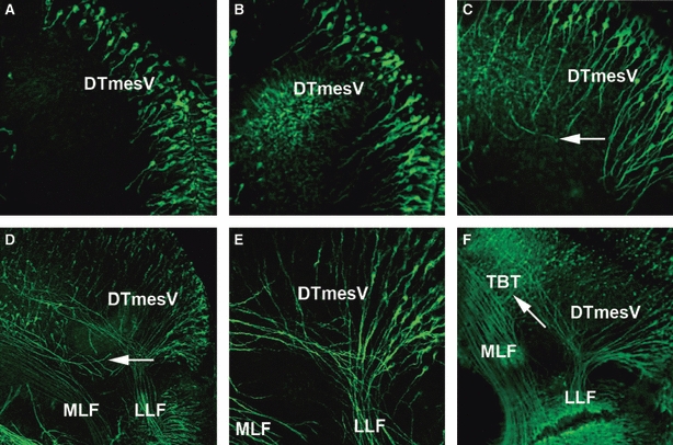
Fig. 6.

Formation of the DTmesV in the dorsal mesencephalon. (A) HH14: neurones have differentiated along the dorsal midline of the mesencephalon and have begun projecting their axons ventrally into the mesencephalon. (B) HH15: the axons of the DTmesV have projected further into the alar plate of the mesencephalon. (C) HH16: DTmesV axons have started turning caudally as indicated with arrow. (D) HH17: the caudally projecting DTmesV axons have formed a dense bundle (LLF) in the alar plate. Some MLF axons project from the MLF towards the DTmesV (arrow). (E) HH17: high magnification of the MRB, showing the LLF projecting as a compact bundle into the rhombencephalon. (F) HH18: LLF and MLF are well separated, and no axons crossing between them are detected. In the mesencephalon, tecto-bulbar axons project perpendicular to LLF and MLF towards the ventral midline (arrow). DTmesV, descending tract of the mesencephalic nucleus of the trigeminal nerve; LLF, lateral longitudinal fascicle; MLF, medial longitudinal fascicle; TBT, tectobulbar tract.
