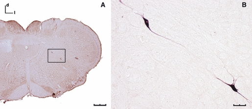
Fig. 4.

Coronal sections of the medulla oblongata showing aberrant neurons labelled following the application of BDA to the SLN (DAB staining, bright-field microscopy). (A) Two aberrant neurons in an intermediate location between the Amb and the 10N (scale bar: 500 μm). (B) Enlargement of the box in (A), both aberrant neurons show fusiform morphology (scale bar: 50 μm). d, dorsal; l, lateral.
