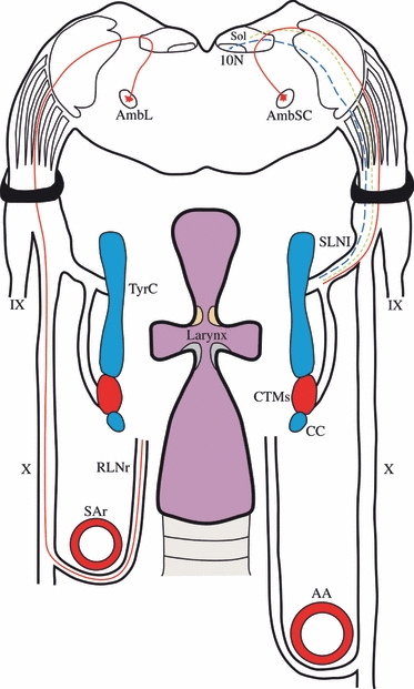
Fig. 6.

Schematic drawing summarising the fibres contained in the SLN and RLN of the rat. The SLN has been represented on the right side of the picture, whereas the RLN is on the left. The SLN contains special visceral efferent fibres (red-continuous line), general visceral efferent fibres (blue-dashed line) and general visceral afferent fibres (green-dotted line). The RLN only has special visceral efferent fibres (red-continuous line). 10N, dorsal motor nucleus of the vagus; AA, aorta artery; AmbL, loose formation of the nucleus ambiguus; AmbSC, semicompact formation of the nucleus ambiguus; CC, cricoid cartilage; CTMs, cricothyroid muscle; IX, glossopharyngeal nerve; RLNr, right recurrent laryngeal nerve; SAr, right subclavian artery; SLNl, left superior laryngeal nerve; Sol, nucleus of the solitary tract; TyrC, thyroid cartilage; X, vagus nerve.
