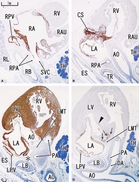
Fig. 1.

Sagittal sections of a 9-week human fetus. Panel A (D) is the most rightward (leftward) in the figure. Right side corresponds to the ventral side of the body. Intervals between panels are 0.6 mm (A–B), 0.5 mm (B–C) and 0.2 mm (C–D), respectively. Panels A, B and D represent desmin expression; panel C represents MHC. Desmin reactivity is seen along the atrium (LA, RA), proximal parts of the pulmonary vein (LPV, RPV), superior vena cava (SVC) and cavernous sinus (CS) at and around the atrioventricular node (arrows in panel D). The auricle is almost negative. MHC is positive in the ventricle as well as in the atrium. Asterisk in panel C indicates a protrusion of the left atrium. DA, ductus arteriosus (Botallo's duct). All panels were prepared at the same magnification (scale bar in panel A). TH, thymus.
