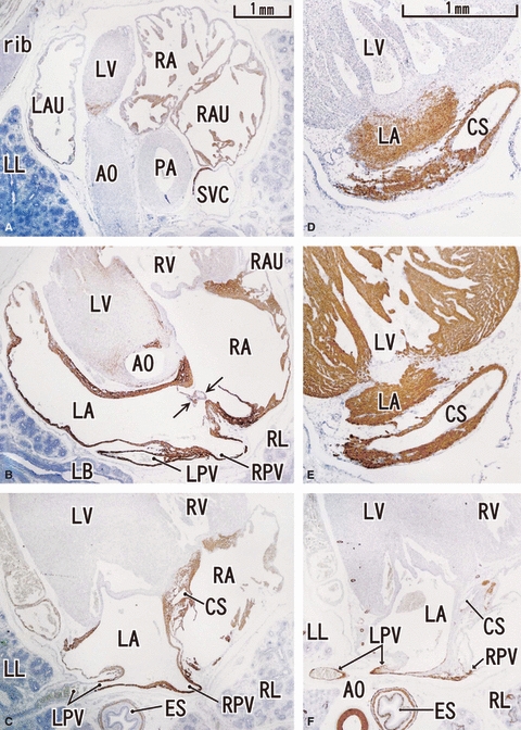
Fig. 2.

Horizontal sections of a 12-week human fetus. Panel A (F) is the most cranial (caudal) in the figure. Upper part corresponds to the ventral side of the body. Intervals between panels are 1.2 mm (A–B), 1.0 mm (B–C) and 1.2 mm (C–F), respectively. Panels D and E are magnified sections in the intermediate levels between panels C and F. Panels A–D represent desmin, panel E is MHC and panel F is α-SMA. Desmin expression is seen along the atrium (LA, RA), proximal parts of the pulmonary vein (LPV, RPV), superior vena cava (SVC) and the cavernous sinus (CS). The auricle is weakly positive. MHC (panel E) is positive in the ventricle as well as in the atrium. α-SMA (panel F) is positive along the vascular wall. Arrows in panel B indicate a valve-like structure between the atria. Panels A–C and F (D and E) were prepared at the same magnification (scale bars in panels A and D).
