
Fig. 3.
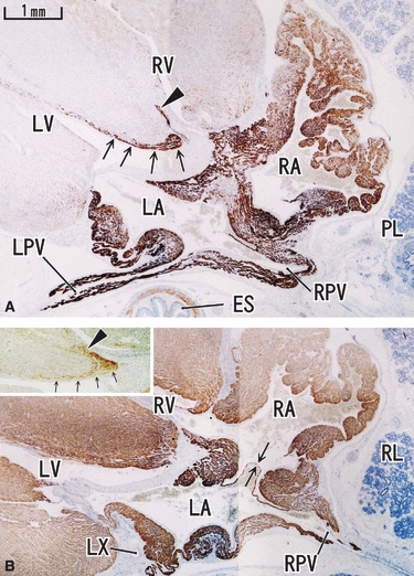
Horizontal section of an 18-week human fetus. Panel A (desmin) is located 5 mm superior to panel B (MHC). Upper part corresponds to the ventral side of the body. Proximal parts of the pulmonary vein (LPV, RPV) are positive for desmin, but the reactivity disappears suddenly in the distal parts (panel A). The auricle around the right atrium (RA) is also strongly positive for desmin. Arrows and arrowhead in panel A indicate the desmin-positive conduction system. These structures with labels are also seen in the insert in panel B (section near panel A is stained for NSE). Arrows in panel B indicate a valve-like structure between the atria. All panels and inserts were prepared at the same magnification (scale bar in panel A). LX, left circumferential branch of the coronary artery.