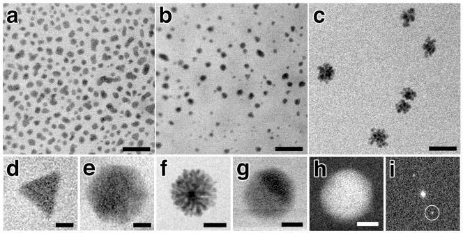
Figure 3. Nanoparticle growth under varying chemical composition of parent solution.
In situ Bright Field Scanning TEM images of 2:1 (a), 1:1 (b) and 1:1.25 (c) Pb:S solutions each at 5,000x dilution. Panels (d–g) show a gallery of differently shaped nanoparticles observed in situ including trigonal (d), hexagonal (e) flower-like (f), and spherical (g). Dark Field Scanning TEM image (h) of the same nanoparticle in (g) and the corresponding FFT (i). Lattice fringes for the (220) plane of PbS at 0.21 nm resolution can be seen in (g&h) and Bragg reflections circled in (i). Scale bars represent 100 nm (a–c), 12.5 nm (d&e), 25 nm (f), and 2.5 nm (g&h).

