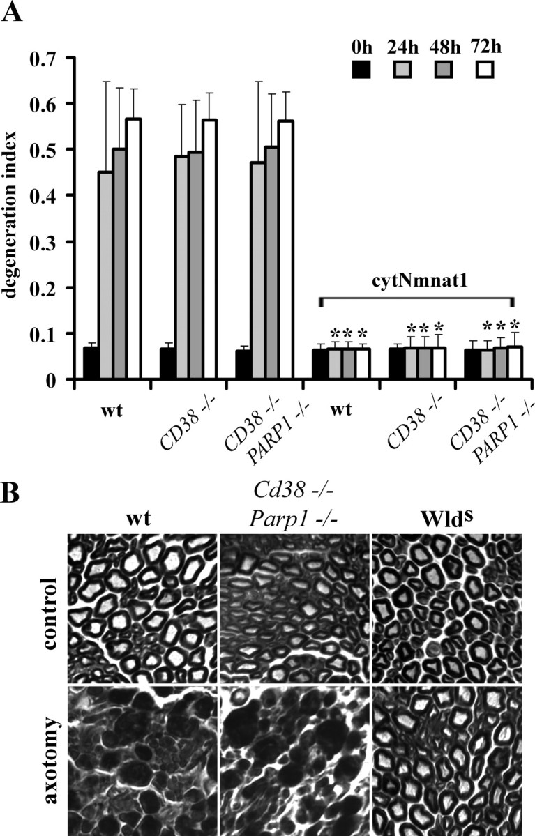
Figure 4.

Axonal degeneration proceeds normally in Cd38−/−Parp1−/− mice despite high NAD+ levels. A, In vitro axonal degeneration assays were performed using DRG neurons from mice with the indicated genotypes infected with lentivirus expressing cytNmnat1 or EGFP. The DI was calculated as outlined in Materials and Methods. Sixteen fields were evaluated for each condition, and each experiment was repeated three times. The degeneration index value ± SD at 0, 24, 48, and 72 h is displayed. *Significant difference (p < 0.001) between neurons expressing cytNmnat1 and those expressing EGFP of the indicated genotypes. B, Sciatic nerves in mice of the indicated genotypes were transected, and the distal segments were harvested 7 d later. Transverse sections of the distal segments stained with toluidine blue are displayed. While axons from wlds mice are preserved, degenerating axon profiles are abundant in nerves from Cd38−/−Parp1−/− and wild-type (wt) mice.
