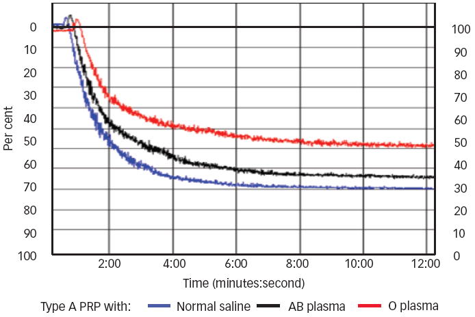
Figure 1. Typical Platelet Aggregation of a Type A Donor.

500μl of platelet-rich plasma (PRP) of platelet (PLT) type A was tested against 20μM of adenosine diphosphate (ADP) after 10 minutes of incubation at 37°C with 50μl of normal saline (blue line: aggregation of 81%), AB plasma (black line: aggregation of 75%), or O plasma (red line: aggregation of 58%, 29% inhibition).
