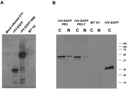
Figure 2.
WB analysis of recombinant PR3 and HNE expression. CV1 cells were infected with rVVs expressing HNE, PR3, and PR3-T fused to EGFP. Cytoplasmic (C) and nuclear (N) fractions were prepared 24 hours after infection with rVVs at an MOI of 1. The blot was probed with an Ab to HNE (A) or to EGFP (B). Apparent molecular weights are indicated in kilodaltons. This image is representative of 3 rVV infection experiments.

