Figure 6.
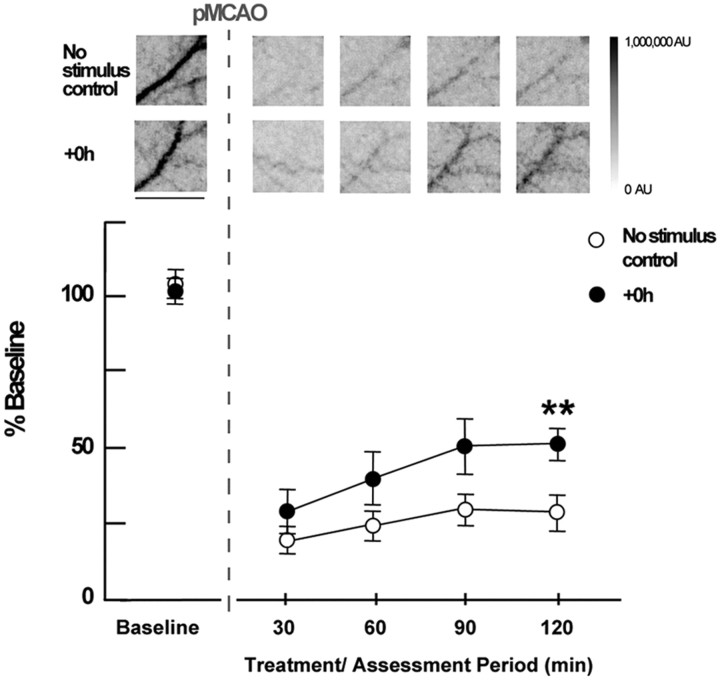
LSI experiments demonstrate that post-pMCAO blood flow return in MCA is induced by whisker stimulation treatment. Insets, Representative cases of no-stimulus control and +0 h animals presented as linearly gray-scaled LSI images taken at baseline and following pMCAO during treatment at ∼30 min intervals. Scale bar, 1 mm. The dark vessel diagonally traversing the image in each case is a cortical branch of MCA (most clear in preocclusion baseline images; other vessels such as dural vessels are also visible, but much less apparent than MCA at baseline). Representative segments of MCA branch ROIs above the region of somatosensory cortex, distal to pMCAO are shown [(for an in depth description regarding criteria for MCA regions of interest analyzed, see the Methods section of Lay et al. (2010)]. Means and SEs for MCA for no-stimulus and +0 h animals at baseline, following pMCAO, and during whisker stimulation. **p < 0.01, significant increase in +0 h flow compared with the no-stimulus control group.

