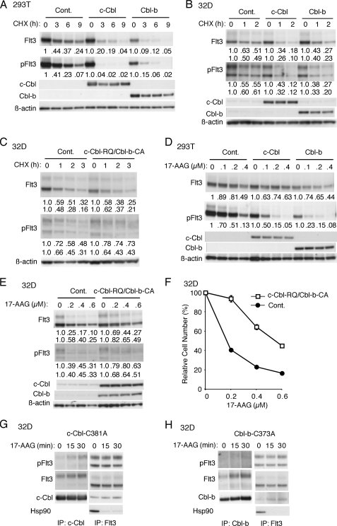
FIGURE 5.
c-Cbl and Cbl-b play important roles in constitutive and 17-AAG-induced degradation of autophosphorylated Flt3-ITD. A, 293T cells were transfected on 6-well plates with 0.5 μg each of pCEFL (Cont.), pCEFL-c-Cbl (c-Cbl), or pCEFL-Cbl-b (Cbl-b) as indicated and 0.5 μg of pcDNA3-Flt3-ITD. Two days after transfection cells were treated with 100 μg/ml CHX for the indicated times and harvested. Total cell lysates were analyzed by immunoblot analysis using anti-Flt3 followed by reprobing with anti-phospho-Tyr-591-Flt3 (pFlt3), anti-c-Cbl, anti-Cbl-b, and anti-β-actin as indicated. Relative expression levels of Flt3-ITD and Flt3-ITD phosphorylated on Tyr-591 were determined by densitometric analysis of the anti-Flt3 and anti-pFlt3 data, respectively, and are shown below each panel. B, after culture with 1 μg/ml DOX for 24 h, Ton.32D/Flt3-ITD/pRevTRE (Cont.), Ton.32D/Flt3-ITD/pRevTRE-c-Cbl (c-Cbl), and Ton.32D/Flt3-ITD/pRevTRE-Cbl-b (Cbl-b) cells were treated with 25 μg/ml CHX for the indicated times and lysed. Cells lysates were subjected to Western blot analysis. Relative expression levels of the mature (upper rows) and immature (lower rows) forms of Flt3-ITD and Flt3-ITD phosphorylated on Tyr-591 were determined by densitometric analysis of the anti-Flt3 and anti-pFlt3 data, respectively, and are shown below each panel. C, after culture with DOX, Ton.32D/Flt3-ITD (Cont.) and Ton.32D/Flt3-ITD/c-Cbl-R420Q/Cbl-b-C373A (c-Cbl-RQ/Cbl-b-CA) cells were treated with 10 μg/ml CHX for the indicated times, and cell lysates were analyzed. D, 293T cells were transfected on 6-well plates with 0.4 μg each of pCEFL (Cont.), pCEFL-c-Cbl (c-Cbl), or pCEFL-Cbl-b (Cbl-b) as indicated and 0.4 μg of pcDNA3-Flt3-ITD. Two days after transfection cells were treated for 5 h with the indicated concentrations of 17-AAG and harvested. Total cell lysates were analyzed by immunoblot analysis. E, after culture with DOX, Ton.32D/Flt3-ITD (Cont.) and Ton.32D/Flt3-ITD/c-Cbl-R420Q/Cbl-b-C373A (c-Cbl-RQ/Cbl-b-CA) cells were treated for 2 h with indicated concentrations of 17-AAG, and cell lysates were analyzed. F, after culture with DOX, Ton.32D/Flt3-ITD (Cont.) and Ton.32D/Flt3-ITD/c-Cbl-R420Q/Cbl-b-C373A (c-Cbl-RQ/Cbl-b-CA) cells were further cultured for 36 h with the indicated concentrations of 17-AAG and 1 μg/ml DOX in the absence of IL-3. Numbers of viable cells were measured by the XTT assay and are expressed as percentages of the cell number without 17-AAG. Each data point represents the mean of triplicate determinations, with error bars indicating S.E. G and H, Ton.32D/Flt3-ITD/pRevTRE-c-Cbl-C381A (G) or -Cbl-b-C373A (H) cells cultured with DOX were pretreated with 5 μm MG132 for 30 min and then treated with 0.5 μm 17-AAG for the indicated times. Cells were lysed and subjected to immunoprecipitation with anti-c-Cbl, anti-Cbl-b, or anti-Flt3, as indicated and analyzed with immunoblot analysis using indicated antibodies.

