FIGURE 5.
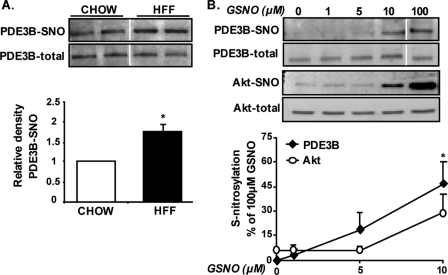
PDE3B is a potential target of S-nitrosylation in adipocytes. A, degree of S-nitrosylation of PDE3B was determined in adipose tissue of HFF obese or chow-fed lean control mice using the biotin-switch assay, with several specific optimizations, as detailed under “Experimental Procedures.” Shown are representative blots and densitometry analysis of results obtained from three chow-fed and six HFF mice, p = 0.04. B, lysates of differentiated epididymal pre-adipocyte cell line were exposed to 0, 1, 5, 10, or 100 μm GSNO for 30 min at room temperature, after which S-nitrosylations of PDE3B and Akt were assessed. Densitometry of PDE3B-SNO and Akt-SNO per their respective total protein is derived from five independent experiments and presented as the percent of intensity achieved with 100 μm GSNO. Vertical white line denotes splicing of the same membrane for clearer presentation.

