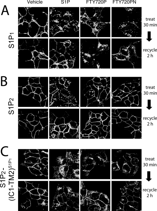
FIGURE 5.
Receptor internalization of S1P1-, S1P2-, and S1P2(IC1-TM2)S1P1-GFP. Stably transfected HEK293 cells expressing WT S1P1 (A), WT S1P2 (B), or S1P2(IC1-TM2)S1P1 (C) GFP-fusion proteins were exposed to a 100 nm concentration of either vehicle (BSA), S1P, FTY720P, or FTY720PN for 30 min. Cells were rinsed and incubated for 2 h in the presence of cycloheximide to allow receptor recycling in the absence of new protein synthesis. The cells were fixed, and receptor localization was examined by laser-scanning confocal fluorescence microscopy.

