FIGURE 1.
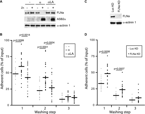
ASB2α-induced FLNa degradation enhances adhesion of hematopoietic cells to fibronectin. PLB985/MT-FLAG (−), PLB985/MT-ASB2α (α), and PLB985/MT-ASB2αLA (α LA) cells were cultured for 16 h without or with 60 μm ZnSO4 (A and B), loaded with calcein AM, serum arrested for 30 min, and plated on fibronectin-coated wells for 10 min in the presence of 1 mm Mn2+ (B). A, ASB2α and FLNa expression in cells cultured in the absence (−) or presence (+) of ZnSO4. Protein extracts (10 μg) were separated by SDS-PAGE and immunoblotted for FLNa, ASB2α, and α-actinin 1 as loading control. B, adhesion of PLB985/MT-FLAG, PLB985/MT-ASB2α, and PLB985/MT-ASB2αLA cells to fibronectin was assayed following the first, second, and the third washing steps with PBS. Dot plots show the overall distribution, the line shows the median value. p values were calculated using the Mann-Whitney t test. (Sample size: PLB985/MT-FLAG = 21, PLB985/MT-ASB2α = 27, and PLB985/MT-ASB2αLA = 27; from 7, 9, and 9 independent experiments, respectively.) C, FLNa expression in PLB985 FLNaKD and LucKD cells. Protein extracts (10 μg) were separated by SDS-PAGE and immunoblotted for FLNa and α-actinin 1. D, adhesion of PLB985 FLNaKD and PLB985 LucKD cells. Adhesion assays were performed as described above (sample size: PLB985 FLNaKD = 18 and PLB985 LucKD = 18; from 6 independent experiments).

