FIGURE 6.
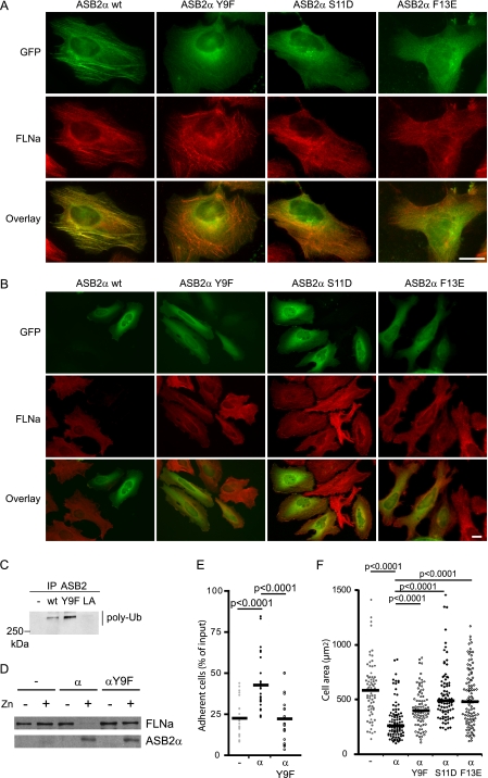
ASB2α Tyr-9, Ser-11, and Phe-13 are required for ASB2α to target FLNa. HeLa cells were transfected with GFP-ASB2α, GFP-ASB2αY9F, GFP-ASB2αS11D, and GFP-ASB2αF13E expression vectors and analyzed 8 (A) and 48 h (B) after transfection using an antibody against FLNa. A, ASB2α Tyr-9, Ser-11, and Phe-13 are required for co-localization of ASB2α and FLNa onto stress fibers. B, ASB2α-induced FLNa degradation in HeLa cells is dependent on Tyr-9, Ser-11, and Phe-13 of ASB2α. Scale bar represents 20 μm. C, ASB2αY9F activates formation of polyubiquitin chains by the E2 enzyme. EloB, EloC, Cul5, and Rbx2 together with wild-type ASB2α, ASB2αLA, and ASB2αY9F were expressed in Sf21 cells. ASB2α complexes were immunoprecipitated (IP) with anti-ASB2 antibodies, and incubated with Uba1, Ubc5a, ubiquitin, and ATP. Their ability to stimulate polyubiquitylation was assessed by Western blotting with antibodies to polyubiquitylated proteins. D and E, effects of ASB2αY9F on adhesion of hematopoietic cells to fibronectin. PLB985/MT-FLAG (−), PLB985/MT-ASB2α (α), and PLB985/MT-ASB2αY9F (αY9F) cells were cultured for 16 h without or with 60 μm ZnSO4 (D and E), loaded with calcein AM, serum arrested for 30 min, and plated on fibronectin-coated wells for 10 min in the presence of 1 mm Mn2+. D, 10-μg aliquots of protein extracts of cells cultured in the absence (−) or presence (+) of ZnSO4 were immunoblotted with antibodies to ASB2α and FLNa. E, adhesion of PLB985/MT-FLAG, PLB985/MT-ASB2α, and PLB985/MT-ASB2αY9F cells to fibronectin was assayed following the second washing step with PBS. Dot plots show the overall distribution, the line shows the median value. (Sample size: PLB985/MT-FLAG = 21, PLB985/MT-ASB2α = 27, and PLB985/MT-ASB2αY9F = 21; from 7, 9, and 7 independent experiments, respectively.) F, effects of ASB2αY9F, ASB2αS11D, and ASB2αF13E on cell spreading on fibronectin of NIH3T3 cells. NIH3T3 cells were transfected with GFP (−), GFP-ASB2α (α), GFP-ASB2αY9F (αY9F), GFP-ASB2αS11D (αS11D), or GFP-ASB2αF13E (αF13E) expression vectors for 24 h, trypsinized, and serum arrested for 1 h in suspension. Cells were plated on fibronectin-coated coverslips and fixed after 45 min. Cell areas of at least 100 cells from three independent experiments were measured. Dot plots show the overall distribution, lines show the median values. p values were calculated using the Mann-Whitney t test.

