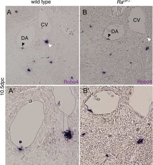
FIGURE 1.
Sox18 knock-out mice show polar loss of Robo4 expression in caudal vein (CV). A and B are transverse section of Robo4 in situ 10.5 days postcoitus (dpc) embryos (WT and RaOp−/−). The expression of Robo4 is detected in the endothelium of both DA and caudal vein in dorsolateral polarized fashion. The black arrowhead shows Robo4 expression in the DA, and the white arrowhead shows the expression in the caudal vein. A′ and B′ are, respectively, high power images of the A and B, respectively, with the dotted line outlining the DA and CV. ISH was performed on 3 WT and 3 RaOp−/− homozygous embryos.

