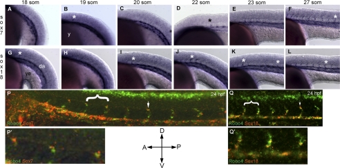
FIGURE 2.
Montage of robo4, sox7, and sox18 endogenous expression across embryonic zebrafish development. Whole mount for sox7 (A–F), and sox18 (G–L) ISH embryos were performed as indicated under “Experimental Procedures.” Embryos were positioned with anterior (A) to the left, posterior (P) to the right and dorsal (D) to the top and ventral (V) to the bottom as indicated by the orientation bars. Embryos were staged according to the somite numbers as indicated in the respective panels. Asterisks (black and white) indicate ISVs in the zebrafish trunk region. da, dorsal aorta; y, yolk; ye, yolk extension. P and Q are whole mount two-color confocal fluorescent sox7 or sox18 (red) with robo4 (green) ISH images of 24 hpf zebrafish trunk. White arrows indicate ISVs co-localized for robo4 and sox7/18 transcript. P′ and Q′ are higher magnification of regions highlighted by white brackets in P and Q, respectively.

