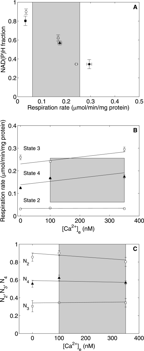
FIGURE 6.
Ca2+ effect on State 2, 3, and 4 respiration and normalized NAD(P)H levels at subsaturating pyruvate/malate concentrations and 2.5 mm initial Pi. A shows a plot of NAD(P)H versus respiration rates at low (open symbols) and high (closed symbols) [Ca2+]e. B shows the respiration data plotted against [Ca2+]e during State 2 (○), State 3 (□), and State 4 (▴). C shows the NAD(P)H fractions plotted against [Ca2+]e during State 2 (○), State 3 (□), and State 4 (▴). All data are presented as means ± S.D. of three replicates.

