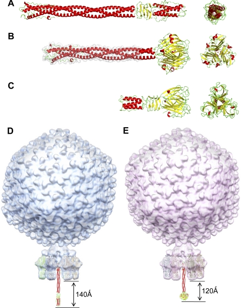
FIGURE 5.
Modeling bacteriophage tail needles. A, crystal structure of P22 tail needle gp26 (PDB code 3C9I). B, homology model of the full-length Sf6 tail needle. The portion of the tail needle helical core missing (residues 1–132) in the construct used for crystallography was modeled using Phyre (38) and is highlighted in gray. C, crystal structure of the receptor binding protein from lactococcal phage TP901-1 base plate (PDB code 3EJC) (20, 39). D, a composite model of bacteriophage P22 and Sf6 (E) mature virions generated by fitting the atomic structure of gp26 tail needles and tailspikes into the asymmetric cryo-EM reconstruction of mature P22 (EMDB ID 1220) (6).

