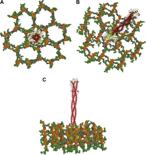
FIGURE 6.
A structural model for Sf6 tail needle penetration of Shigella cell wall. Top (A), tilted (B), and side (C) view of the Sf6 tail needle embedded into a pore of the Gram-negative cell wall, according to the structure of PG determined by Meroueh et al. (49). Glycan strands are colored orange, and the cross-linking stem peptides are colored green, with d-Glu in purple. The Sf6 tail needle is shown as a transparent solvent-accessible gray surface overlaid to the ribbon structure.

