Figure 3.
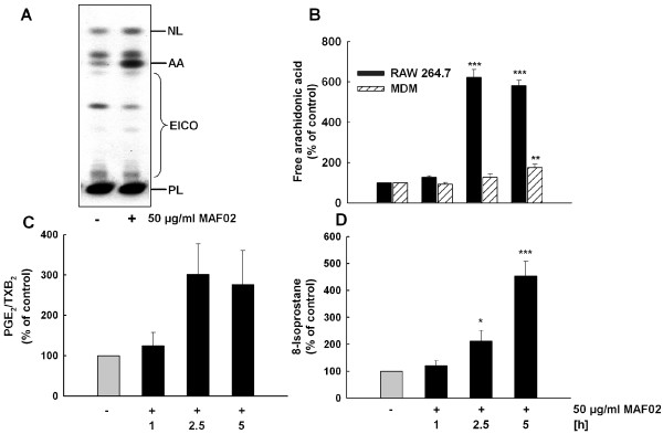
Exposure to fly ash particles induces liberation of AA and release of PGE2 and 8-isoprostane from macrophages. RAW264.7 macrophages and human MDM, pre-labelled with [14C]arachidonic acid, were incubated for 1, 2.5, and 5 h with MAF02 particles at 50 μg/ml (13.2 μg/cm2). After lipid extraction, AA and lipid mediators PGE2/TXB2 were separated by TLC (A, example of RAW264.7 cells treated for 2.5 h), and pixel images generated by phosphoimaging were quantified by OptiQuant® software. Semiquantitative data on AA (B) and PGE2/TXB2 (C, only RAW264.7) liberation based on electronic autoradiographic values are expressed as percentage of control cells. (D) RAW264.7 macrophages were incubated for 1, 2.5, and 5 h with fly ash particles at 50 μg/ml (10.4 μg/cm2) and 8-isoprostane in the medium was determined by EIA. Results are presented as the mean of three independent experiments and error bars represent the standard error of the mean. Statistically significant differences from untreated control cells (*p < 0.05, **p < 0.01, *** p < 0.001). NL, neutral lipids; AA, free arachidonic acid; EICO, eicosanoids; PL, phospholipids.

