Figure 4.
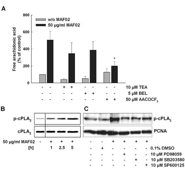
MAF02-induced liberation of arachidonic acid is dependent on cPLA2. (A) RAW264.7 macrophages, pre-labelled with [14C]arachidonic acid, were incubated with AACOCF3 (inhibitor of cPLA2), BEL (inhibitor of iPLA2) or TEA (inhibitor of sPLA2), and subsequently treated with 50 μg/ml MAF02 particles (13.2 μg/cm2) for 2.5 hours. After lipid extraction, the free arachidonic acid and its metabolites were separated by TLC, visualized by autoradiography and the optical density of the bands were analyzed by Odyssey® software. [14C]arachidonic acid liberation data are expressed as percentage of untreated control cells (100%). Results are shown as the mean ± s.e.m. of three independent experiments. *Significantly decreased compared to 50 μg/ml (13.2 μg/cm2) MAF02-treated control cells (*p < 0.05). (B) RAW264.7 macrophages were exposed to 50 μg/ml MAF02 particles (15.6 μg/m2) for 1, 2.5 and 5 hours and whole cell lysates were analyzed for the phosphorylated cPLA2 protein by Western blotting. The respective non-phosphorylated protein was used as loading control. (C) RAW264.7 macrophages were pre-incubated with MAPK inhibitors PD98059, SB203580 and SP600125 (each 10 μM) and then exposed to 50 μg/ml MAF02 particles (15.6 μg/m2) for 5 hours. Whole cell lysates were analyzed for the phosphorylated cPLA2 protein, COX-2 and PCNA as loading control. The shown blots are representative of three independent experiments.

