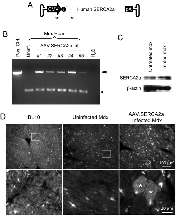
Figure 1.
AAV-9 mediated SERCA2a transduction in the mdx heart. A, Schematic outline of the AAV SERCA2a vector used in the study. The human SERCA2a cDNA is driven by the CMV promoter. i, intron. Arrows mark the locations of the PCR primers. B, PCR detection of the AAV SERCA2a vector genome in the mdx heart. Pos. Ctrl., the SERCA2a cis plasmid; Uninf., from an uninfected mdx heart; #1 to #5, from five AAV-9 SERCA2a vector infected mdx mouse hearts. Each line represents PCR result from one mouse; H2O, no DNA was added in the PCR reaction. Arrowhead, the 519 bp diagnostic band for the AAV SERCA2a genome; Arrow, the 160 bp diagnostic band for the CFTR gene (internal control). C, Representative SERCA2a western blot. β-actin was used as the loading control. D, Representative SERCA2a immunofluorescence staining images from BL10, mdx and AAV-9 SERCA2a infected mdx hearts. Enlarged images (bottom panels) are the boxed areas from the corresponding low-power photomicrographs (top panels). Asterisk, AAV SERCA2a transduced cardiomyocytes.

