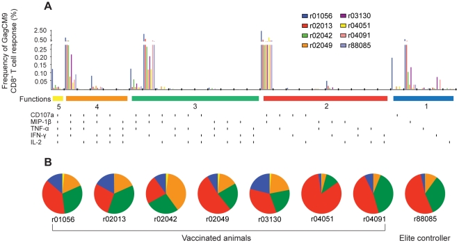
Figure 2. Multifunctional GagCM9-specific CD8+ T cell responses.
(a, b) Using intracellular cytokine staining, we analyzed the ability of GagCM9-specific peptide-stimulated PBMC to degranulate (CD107a) and secrete MIP-1β, TNF-α, IFN-γ, and/or IL-2 at week ten post-infection. (a) Bar graphs indicate the GagCM9-specific CD8+ T cell response frequency for each molecule alone or in combination with the other tested molecules from each animal. Each vertical black line below the horizontal colored bars indicates positivity for CD107a, MIP-1β, TNF-α, IFN-γ and/or IL-2. (b) Pie graphs indicate the percentage of GagCM9-specific CD8+ T cells that had five molecules (yellow), four molecules (orange), three molecules (green), two molecules (red) or one molecule (blue). Animal r88085 was a Mamu-A*01+ delta-nef-vaccinated, SIVsmE660-infected elite controller from a previous study who mounted a GagCM9-specific CD8+ T cell response.

