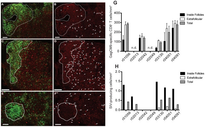
Figure 4. GagCM9-specific CD8+ T cells reside in the lymph nodes of infected animals at high effector to target ratios.
Representative images of Mamu A*01-restricted GagCM9-specific tetramer positive CD8+ T cells (red) and CD20+ B cells (green) in lymph node sections taken during the chronic phase of infection from animals r03130 (a, b), r04051 (e, f), and r04091 (c, d). B cell follicles are delineated with a white line. The images to the right (b, d, f) show the same field as presented on the left (a, c, e) with only the red tetramer stain shown. Each tetramer-binding cell is indicated with a white arrow. (g) The frequency of GagCM9-specific tetramer positive CD8+ T cells per mm2 inside and outside of the B cell follicle as well as total tissue was calculated for each animal. (h) The frequency of SIV producing cells per mm2 inside and outside the B cell follicle as well as total tissue was calculated for each animal. Animals r02042 and r02049 did not have any positive cells in the sections examined.

