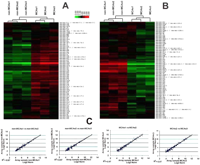
Figure 1. MicroRNA expression in SVZ neural progenitor cells.
Hierarchical clustering of differentially expressed miRNAs (A, B). The data were from 6 individual microarrays (3 arrays per group). The individual expression signal of each miRNA in each array was clustered. The dendrograms (tree diagrams) show the grouping of miRNAs according to the order in which they were joined during the clustering. The color code in the heat maps is linear with green as the lowest and red as the highest. The miRNAs with increased expression are shown in red (A), whereas the miRNAs with decreased expression are shown in green (B). Correlation of the hybridization signal intensities of all the expressed miRNAs among three non-MCAo samples and MCAo showed few differences(C).

