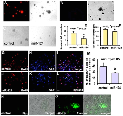
Figure 4. The effect of miR-124a on proliferation and differentiation of neural progenitor cells.
Microscopic images acquired under fluorescent and bright fields after introduction of miRNA mimic indicator conjugated with Dy547 show more than 90% SVZ neural progenitor cells exhibiting red fluorescence, indicating robust delivery efficiency (A). No red fluorescence was observed in SVZ neural progenitor cells in the presence of miRNA mimic indicator but without nanoparticles (B). Panels C and D show neurospheres cultured in the proliferation medium after introduction of miR mimic controls (C) and miR-124a mimics (D), while panels E and F show quantitative data of number (E) and size (F) of neurospheres delivered with mimic controls (control) or miR-124a mimics (miR-124a) in the proliferation medium. Panels G to M show BrdU immunoreactive cells after transfection with miRNA mimic controls (G to I, and M) and miR-124a mimics (J to L, and M). Panels N and O show DCX-EGFP SVZ cells cultured in the differentiation medium after introduction of miRNA mimic controls (N) and miR-124a mimics (O). Scale bar = 100 µm in D and 20 µm in L and O.

