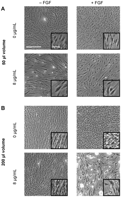
Figure 4. Phase contrast images of hMSCs cultured in 50 µl or 200 µl of medium per well.
Phase contrast images of hMSCs from Fig. 2 and 3 that were either untreated or treated with to 8 µg/mL of polybrene for 24 hr. Cells were cultured for 14 days in either normal culture conditions (A) or under simulated hypoxic conditions (B). The medium was removed prior to image capture. All images are at the same magnification. The insert shows an increased magnification of the cells. White bar = 500 µm (100 µm in insert).

