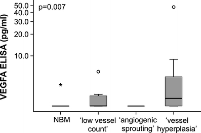
Fig. 4.

Relation between AML derived VEGFA protein levels and vessel morphology in AML at diagnosis. The AML-excreted VEGFA protein level was significantly (p = 0.007) higher in the subgroup with a ‘vessel hyperplasia’ morphology compared with the’angiogenic sprouting’ or the ‘low angiogenic profile’. Box-and-whisker plot limits depict 75th and 25th percentiles and median value (box), and upper/lower quantile ± 1.5 × (interquantile range) (upper and lower whiskers, respectively)
