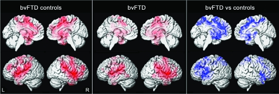
Figure 3. Resting-state fMRI results from the Salience network seed-based analysis of the fronto-insular cortex.
Results are shown only for the behavioral variant frontotemporal dementia (bvFTD) comparisons, since no significant changes in functional connectivity were observed in the asymptomatic MAPT subjects. Left: Patterns of in-phase voxelwise connectivity observed in the bvFTD control subjects. Middle: Patterns of in-phase voxelwise connectivity observed in the subjects with bvFTD. Right: Patterns of reduced (shown in blue) and increased (shown in yellow) in-phase connectivity in the subjects with bvFTD compared to the age-matched controls. Results are shown after cluster-level correction for multiple comparisons at p < 0.05.

