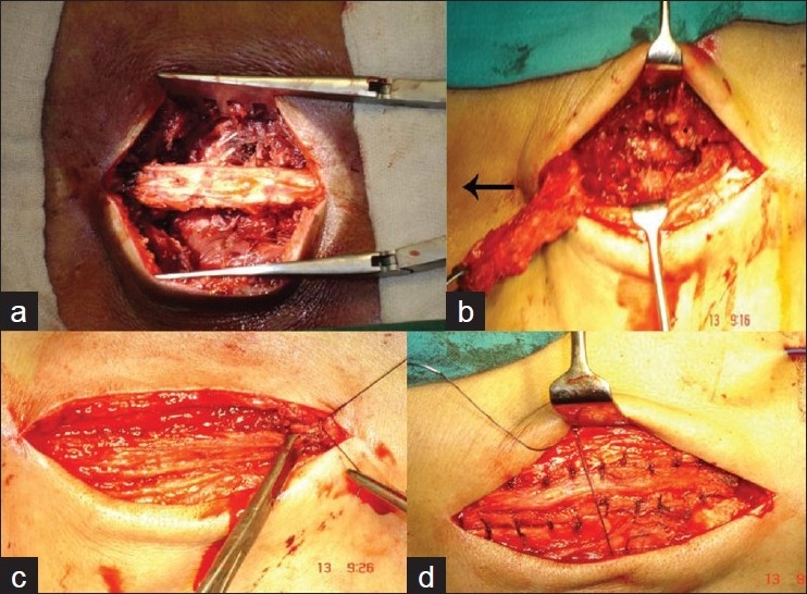
Figure 2.

Intraoperative photographs of the operative steps: (a) median mass of supraspinous, interspinous ligaments and spinous processes exposed; (b) median structures lifted up en masse still attached proximally (arrow); (c) median structures sutured to the distal bed and to paraspinal muscles and aponeurosis; (d) paraspinal muscles are stitched to median structures
