Figure 3.

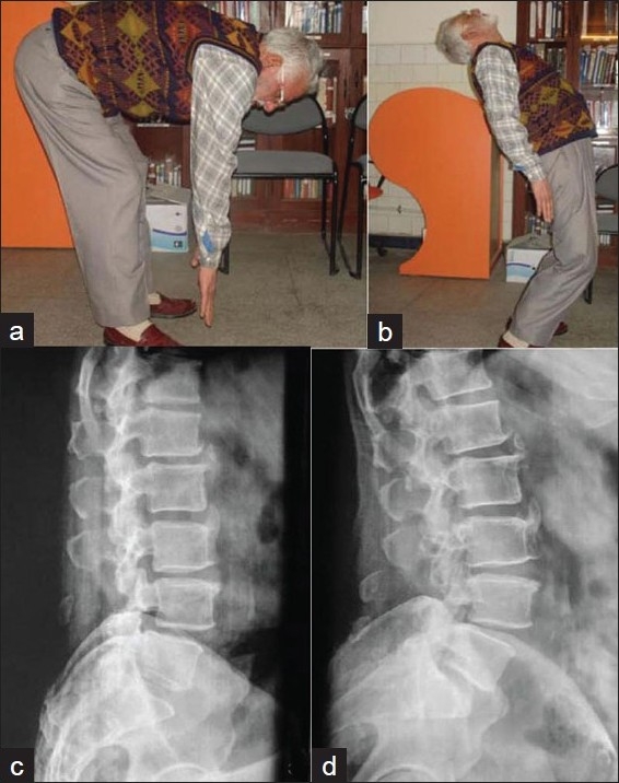
Clinical photographs of a 76 years old patient of degenerative canal stenosis at 6 years followup shows good forward flexion (a) and extension (b). X-ray (lateral view) in flexion (c) and extension (d) shows stable spine

Clinical photographs of a 76 years old patient of degenerative canal stenosis at 6 years followup shows good forward flexion (a) and extension (b). X-ray (lateral view) in flexion (c) and extension (d) shows stable spine